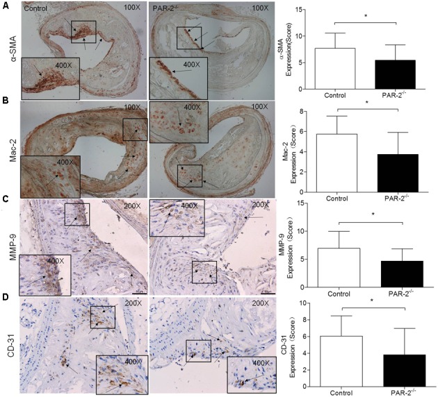
FIGURE 2.

Immunohistochemistry. α-SMA (A) immunostaining for detecting plaque SMC content showed a significant reduction in PAR-2-/- mice, as compared to the controls. Immunohistochemistry staining with antibody against Mac-2 (B), which demonstrates the presence of macrophages within the atherosclerotic lesion, showed a significant reduction in PAR-2-/- mice. Staining for MMP-9 (C) showed a significant reduction in PAR-2-/- mice. Immunohistochemistry staining with antibody against CD-31 (D), which is regarded as evidence of neovascularization, showed a significant reduction in PAR-2-/- mice, as compared to the controls. (C,D) Magnified the shoulder regions of atherosclerotic plaques in the vessel. The arrows show positive stained areas within the atherosclerotic lesions. Data represent mean ± SD. ∗P < 0.05.
