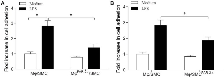
FIGURE 5.
Cell adhesion assay. Mφ was co-cultured with SMC for 15 min in presence (black bars) or absence (white bars) of LPS. LPS induced a significant increase in Mφ adhesion to SMC by approximately threefold compared to unstimulated cells (A). Mφ from PAR-2-deficient mice (MφPAR-2-/-) significantly reduced LPS-induced adhesion (A). SMC from PAR-2-deficient mice (SMCPAR-2-/-) induced a significant inhibitory effect on LPS-induced adhesion (B). Results are presented as fold change of adhesion relative to medium treatment. Data represent mean ± SD. ∗P < 0.05.

