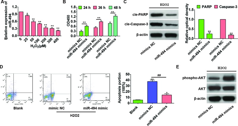
Figure 3. miR-494 reduced the H2O2-induced apoptosis of hepatic AML12 cells by activating AKT in vitro.
(A) Hepatic AML12 cells were treated with H2O2 (0–400 µM) for 6 h, and the level of miR-494 was evaluated by qRT-PCR using U6 as an internal control. *, P<0.05 and **, P<0.01 compared with control cells without H2O2 treatment. (B) AML12 hepatocytes were transfected with mimics NC or miR-494 mimics for 24, 36, and 48 h, and then treated with 200 µM H2O2. Cell viability was measured by CCK8 assay. **, P<0.01 compared with mimics NC group. (C) Apoptosis-associated protein was detected by Western blot. **, P<0.01 compared with mimics NC group. (D) Cell apoptosis rate was detected by flow cytometry (annexin V-FITC staining). *, P<0.05 and **, P<0.01 compared with blank control group. ##, P<0.01 compared with mimics NC group. (E) The activation of AKT was assessed by Western blot. All experiments were performed in triplicate, and data were represented as mean ± S.D.

