Figure 3.

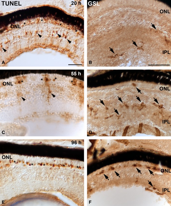
TdT dUTP nick‐end labelling (TUNEL) histochemistry (A,C,E) and Griffonia simplicifolia lectin (GSL) histochemistry (B, D, F) showing the progression of retinal cell death in the photoreceptor layer and changes in distribution pattern and microglial morphology, respectively, in larval teleost after 20 h (A, B), 55 h (C, D) and 96 h (E, F) of light exposure. TUNEL labelling shows that abundant photoreceptors are dying during light treatment (A, C, E) (asterisks). In the first hours of constant light treatment, the number of TUNEL‐positive Müller cells is high (arrowheads in A). However, during the experimental period, cytoplasmic TUNEL staining in the INL became progressively restricted to fewer cells (arrowheads in C, E). In the first hours of constant light treatment, microglial cells are mainly located in the inner border of the INL and in the IPL (arrows in B). As the treatment advances, they progressively become larger and show thicker processes after activation in response to photoreceptor degeneration, and they migrate towards the ONL (arrows in D). Microglial cells invade the photoreceptor layer phagocytosing cell debris (arrows in F). Scale bars: 25 μm (A); 50 μm (B–F). IPL, inner plexiform layer; ONL, outer nuclear layer.
