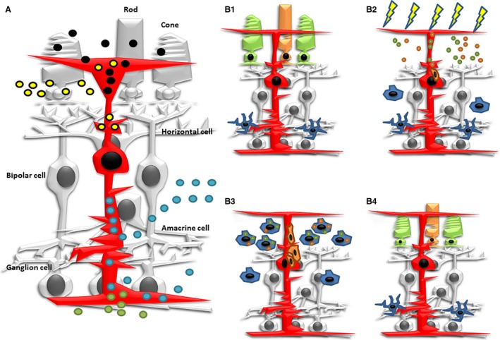
Figure 4.

Schematic diagrams showing several aspects of phagocytosis in Müller glia. (A) Müller cells (in red) phagocytose melanin and outer segment discs (black circles) under physiological conditions. They also engulf photoreceptor cell debris (yellow circles) under pathological and experimental conditions. They participate in the removal of cell debris during development of the retina (blue circles). They also engulf foreign molecules that are injected into the eye (green circles). (B1) Microglial cells in the fish retina are mainly observed in the IPL. (B2) During the first hours of constant intense‐light treatment, cell debris from photoreceptor degeneration is phagocytosed by Müller cells. Signalling molecules activate proliferation activity in phagocytic Müller cells. These signals also activate microglia activation and migration to the photoreceptor layer. (B3) As microglial cells invaded the photoreceptor layer, they become highly phagocytic and participate in the removal of cell debris. However, phagocytic activity of Müller cells progressively decreased. (B4) Migrating precursors from Müller cells proliferate and differentiate into rod and cone photoreceptors, regenerating the missing neurons.
