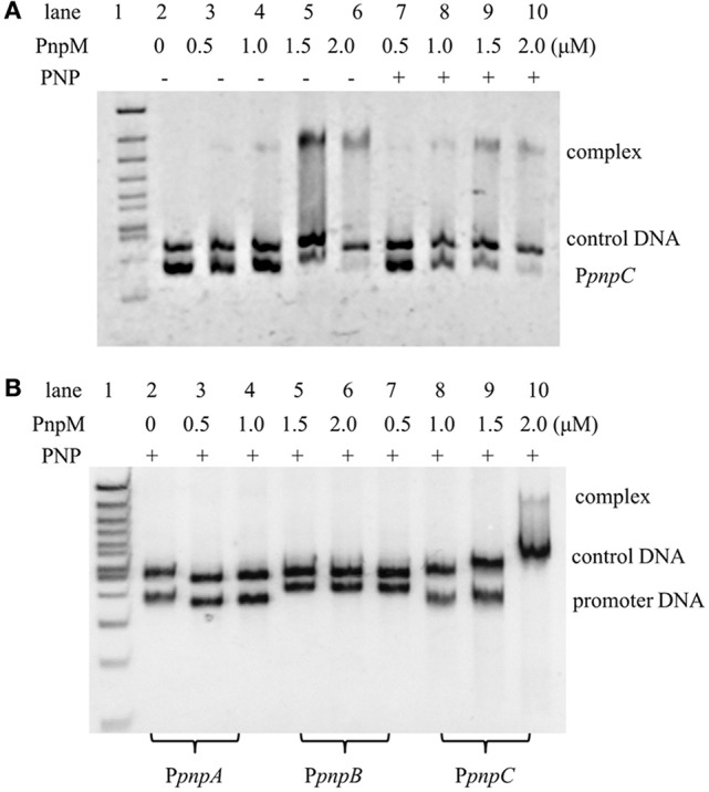
Figure 4.

Electrophoretic mobility shift assays of PnpM binding with pnp promoters. (A) Electrophoretic mobility shift assays of PnpM binding with pnpC promoter (PpnpC). PnpM binding with PpnpC with PNP or without PNP. +, Stands for with PNP, −, stands for without PNP. The first lane was 100 bp ladder Marker, lanes 2–6 contain 0.03μM DNA, with 0μM, 0.5μM, 1.0μM, 1.5μM, and 2.0μM PnpM, respectively; lanes 7–10 contain 0.03μM DNA and 0.3 mM PNP, with 0.5μM, 1.0μM, 1.5μM, and 2.0μM PnpM, respectively. An approximately 400 bp DNA fragment gfp was used as a control DNA (0.03μM). Free probe is a fragment PpnpC of about 270 bp. (B) PnpM binding with pnp promoters in the present of PNP. The first lane was 100 bp ladder Marker, lanes 2–4 contain 0.03μM 283 bp DNA fragment of PpnpA and 0.3 mM PNP, with 0μM, 1.0μM, and 2.0μM PnpM, respectively; lanes 5–7 contain 0.03μM 334 bp DNA fragment of PpnpB and 0.3 mM PNP, with 0μM, 1.0μM, and 2.0μM PnpM, respectively; lanes 8–10 contain 0.03μM 270 bp DNA fragment of PpnpC and 0.3 mM PNP, with 0μM, 1.0μM, and 2.0μM PnpM, respectively. An approximately 400 bp DNA fragment gfp-1 was used as a control DNA (0.03μM).
