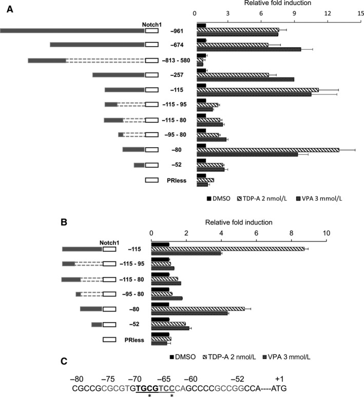
Figure 2.

Deletion analysis of the human Notch1 promoter. BON (A) and H727 (B) cells were treated with TDP‐A and VPA after the cells were transfected with fragments of the Notch1 promoter region joined to a luciferase reporter. A schematic of the individual constructs is shown on the left. The promoter activities of the different deletion fragments were normalized to the relative light unit in cells treated with DMSO vehicle control for each individual constructs. All values were presented as mean relative fold ± SEM. (C) The active DNA sequence of the Notch1 promoter fragment (−80/−1) with the potential transcription factor binding site. The proposed noncanonical AP‐1 binding site is bolded and underlined. Sequences that are different from the canonical AP‐1 binding site are denoted with *.
