. 2017 May 2;8(7):4896–4903. doi: 10.1039/c7sc00700k

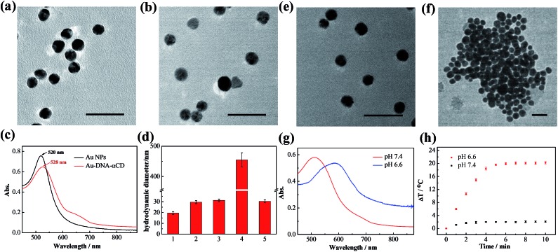
Fig. 1. Characterization of the gold nanomachines and in vitro verification of the fabrication of the gold nanomachines triggered by the mildly acidic environment. High-resolution transmission electron microscopy images of (a) AuNPs and (b) Au-DNA-αCDs. Scale bar represents 50 nm. (c) Absorption spectra of AuNPs and Au-DNA-αCDs. An 8 nm red-shift is apparent after DNA modification and α-CD capping. (d) Hydrodynamic diameters of different samples measured by DLS: (1) AuNPs; (2) Au-DNA-αCD; (3) and (4) Au-DNA-αCDs at pH 7.4 or 6.6, respectively, in PBS buffer (10 mM, Na+ (10 mM) and Mg2+ (5 mM)); (5) Au-DNA-αCDs in PBS buffer (10 mM, Na+ (10 mM), Mg2+ (5 mM), Ca2+(0.5 mM) and DNase I (2 U L–1)) at pH 7.4. (e and f) High-resolution transmission electron microscopy images of the gold nanomachines incubated at pH 7.4 and 6.6 in PBS buffer. Scale bar represents 50 nm. (g) Absorption spectra of gold nanomachines at pH 7.4 and 6.6. The absorption spectrum of the gold aggregates shows a substantial red-shift and possesses a wide range of NIR absorption. (h) Temperature elevation of the gold nanomachines at pH 6.6 or 7.4 as a function of irradiation time. The temperature of the gold nanomachines at pH 6.6 reached 45 °C after irradiation, whereas less than a 3 °C increase was observed at pH 7.4.

Fig. 1