. 2017 May 2;8(7):4896–4903. doi: 10.1039/c7sc00700k

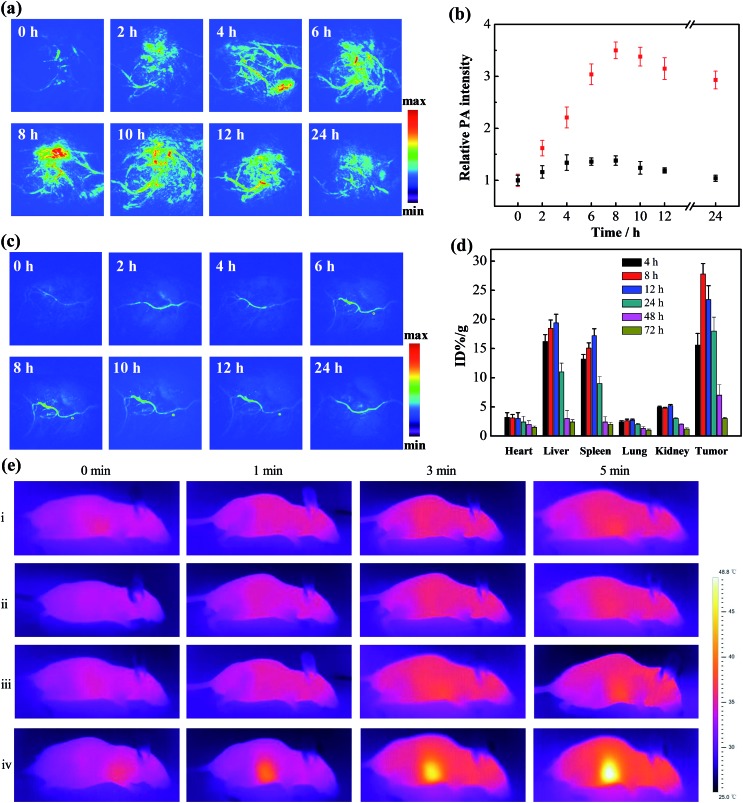
Fig. 3. In vivo PA imaging, biodistribution and photothermal assays. PA images of mice bearing MCF-7 tumors treated with (a) the gold nanomachines or (c) Au-DNA1s at different times (0, 2, 4, 6, 8, 10, 12 and 24 h). (b) Quantification of the PA intensity values in figures (a) (red) and (c) (black) at the corresponding times. (d) Biodistribution of the gold nanoparticles in the different organs of mice (n ≥ 5) treated with the gold nanomachines at different times (4, 8, 12, 24, 48 and 72 h). (e) Photothermal images of mice bearing MCF-7 tumors with different treatments: (i) laser only; (ii) Au-DNA1-αCD ×2 + laser; (iii) Au-DNA1 and Au-DNA2 + laser; (iv) gold nanomachines + laser.

Fig. 3