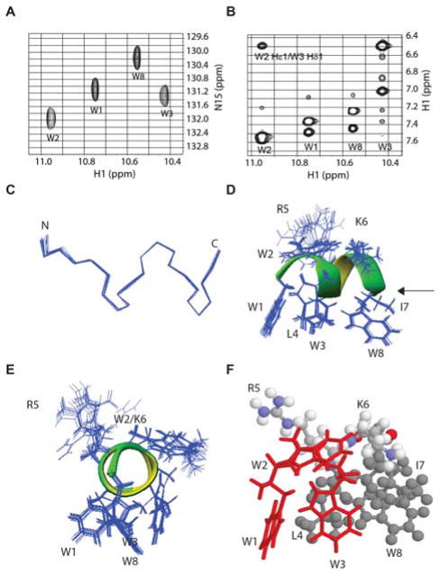
Figure 1.
NMR spectra (A–B) and 3D structure (C–F) of TetraF2W-RK bound to perdeuterated dodecylphosphocholine micelles at 25°C and pH 6.2. Portions of natural abundance 1H-15N HSQC (A) and 1H-1H NOESY (B) spectra illustrate data quality. Also shown are (C) superimposed backbones of an ensemble of 20 structures that form the shortest two-turn helix as indicated by the dihedral angles on the Ramachandran plot (Figure S2), side (D) and end-on (E) views of the amphipathic helix with superimposed side chains, and a side-view of the amphipathic structure featuring the π configuration of the WWW triplet in red (H). Color code: L4, I7, W8 in grey, R5 and K6 in multiple colors: nitrogen, blue; oxygen, red; carbon, grey; and hydrogen, white. NMR data were collected, processed, and assigned as described in the supporting document.4,7a Additional sample conditions and chemical shifts are provided in Table S1.

