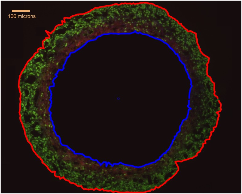
Figure 4.
A DLD1 tumour spheroid, with external boundary marked in red. The oxygen-limited anoxic core (blue outline) is also shown. Green staining is the ki-67 proliferation marker and red is the hypoxia marker EF5. Adapted from Grimes et al with permission from Royal Society Interface.23

