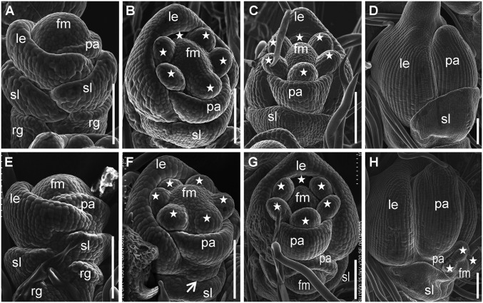
Fig. 2.
Scanning electron micrographs of spikelets at early developmental stages in the WT and lf1. (A–D) WT spikelets. (E–H) lf1 spikelets. (A and E) Stage Sp4. (B and F) Stage Sp5. (C and G) Stage Sp6–Sp7. (D and H) Stage Sp8. Stars indicate stamens. fm, floral meristem; other abbreviations are as in Fig. 1. (Scale bars, 100 µm.)

