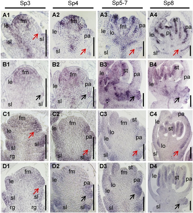
Fig. 4.
Expression pattern of LF1 and miRNA165/166. (A and B) In situ hybridization in the spikelets of the WT (A) and lf1 (B) during stages Sp3–Sp8 using an LF1 antisense probe. (C and D) In situ hybridization in the spikelets of the WT (C) and lf1 (D) during stages Sp3–Sp8 using a miRNA166 complementary locked nucleic acid (LNA)-modified DNA probe. Black arrows in B and D indicate the lateral floral meristem and lateral floret. Red arrows in A and C indicate the axil of the sterile lemma. fm, floral meristem; other abbreviations are as in Fig. 1. (Scale bars, 50 µm.)

