Fig. 7.
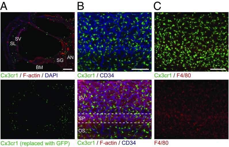
Distribution of resident macrophage-like cells in mouse cochlea. (A) Frozen sections of P4 Cx3cr1GFP/+ cochlea show GFP+ cells distributed in all parts of the cochlea, including the auditory nerve (AN), spiral ganglion (SG), basilar membrane (BM), stria vascularis (SV), and spiral ligament (SL). Sections were counterstained with phalloidin (red) and DAPI (blue) to identify F-actin and nuclei, respectively. (B) Whole-mount preparation of lateral wall of P4 Cx3cr1GFP/+ cochlea stained with CD34 antibody (blue) shows GFP+ cells are mainly located around blood vessels. Anti-CD34 antibody specifically stains vascular endothelium in the inner ear (59). Tissues were counterstained with phalloidin (red). SV, SP, and OS indicate stria vascularis, spiral prominence, and outer sulcus, respectively. (C) Whole-mount preparation of lateral wall of P4 Cx3cr1GFP/+ cochlea stained with F4/80 antibody (red) shows that GFP+ cells are costained with F4/80, indicating these cells have macrophage characteristics. (Scale bars, 100 µm.)

