Fig. 9.
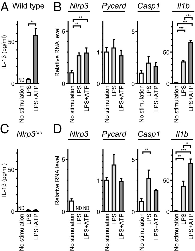
Activation of NLRP3 inflammasome in mouse cochlea. (A) IL-1β levels (mean ± SD) in culture supernatant (total volume = 1.0 mL) from eight wild-type cochleae. Significantly larger amounts of IL-1β were secreted from cultured cochleae in response to LPS+ATP, compared with that in the absence of ATP (unpaired t test, P < 0.01). IL-1β was not detected (ND) in supernatant from cultured cochlea without stimulation. (B) Quantitative RT-PCR analysis of cultured wild-type cochleae. Nlrp3 and Il1b mRNA levels (mean ± SD) were significantly different among each group (one-way ANOVA, **P < 0.01 or ***P < 0.001, respectively). Nlrp3 and Il1b mRNA levels increased in response to LPS compared with levels in the absence of stimulation (Tukey post hoc test: P < 0.01, P < 0.001, respectively). mRNA levels were normalized first to the Actb level and then to the expression level without stimulation. (C) IL-1β levels secreted from cultured Nlrp3∆/∆ cochleae in response to LPS+ATP were not significantly different from those in the absence of ATP. (D) In Nlrp3∆/∆ cochleae, Casp1 and Il1b mRNA levels were significantly different (one-way ANOVA, P < 0.01 or P < 0.001, respectively). Casp1 and Il1b mRNA levels increased in response to LPS compared with levels in the absence of stimulation (Tukey post hoc test: P < 0.001, P < 0.001, respectively). mRNA levels were normalized first to the Actb level and then to the expression level without stimulation of cultured wild-type cochleae.

