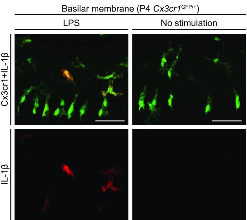
Fig. S7.
Whole-mount preparation of cultured basilar membrane of P4 Cx3cr1GFP/+ mouse cochlea stained with anti–IL-1β antibody. Anti–IL-1β immunoreactivity was detected in a subset of GFP+ cells from cultured cochlea stimulated with LPS, whereas no immunoreactivity was detected in the absence of stimulation. The immunoreactivity reflects intracellular pro–IL-1β because mature IL-1β is secreted. (Scale bars, 50 µm.)

