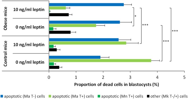
Figure 3.
Proportion of dead cells in blastocysts obtained from the in vitro culture of the two-cell embryos isolated from control and obese mice. According to the presence of particular morphological features [normal nuclear morphology (Mn), nuclear fragmentation or condensation (Ma), nuclear karyolysis (Mk), and positive/negative terminal deoxynucleotidyl transferase dUTP nick end labeling (TUNEL) labeling (T±)], the dead cells were classified as follows: (1) apoptotic, showing fragmented nuclear morphology or TUNEL-positive nucleoplasm or both or (2) other dead cells, showing karyolysis-like nuclear morphology and occasional TUNEL labeling. Embryos from control and obese mice were cultured in leptin-free media or media supplemented with leptin at 10 ng/mL. Numbers of evaluated blastocysts are shown in Table 4. The results are expressed as the mean values + SEM. Asterisks indicate statistical differences between control and obese groups: Kruskal–Wallis test, followed by Dunn’s test, *P < 0.05; ***P < 0.001.

