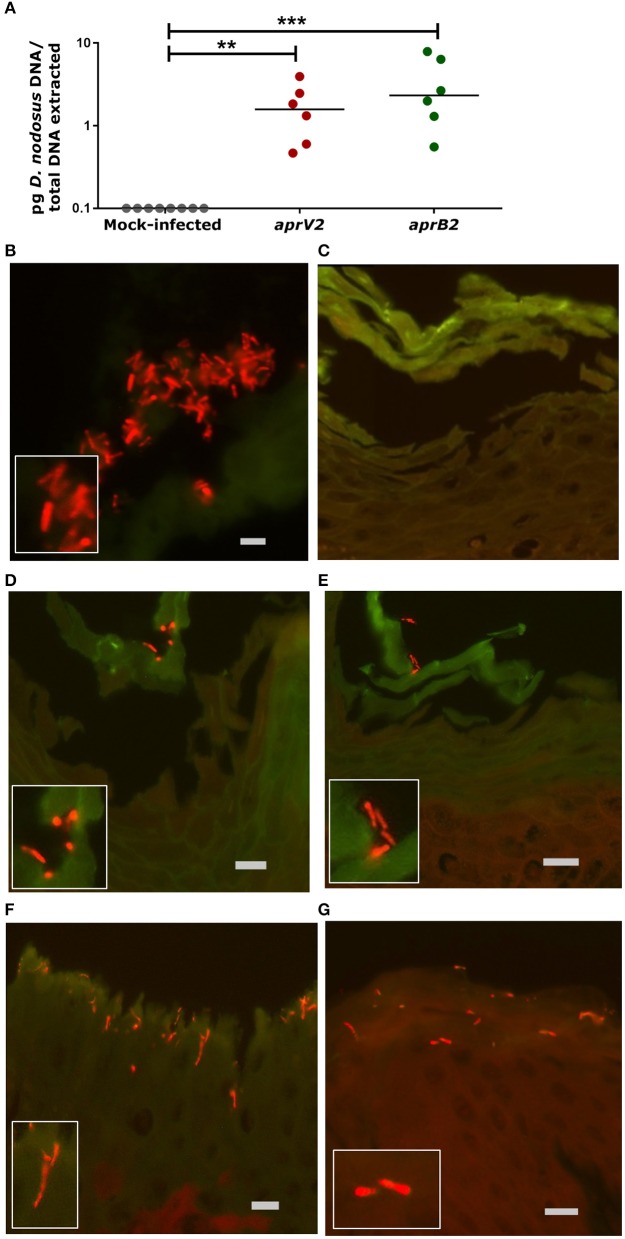
Figure 2.
Infection of the 3D skin explant model with Dichelobacter nodosus. (A) Detection of D. nodosus aprV2 and aprB2 strains in the skin explants using quantitative PCR after 28 h of infection. Each point indicates a single biopsy. 0.1 = results below of the limit of detection. Mean is represented by black bars. Data were analyzed by Dunn's multiple comparisons test using GraphPad Prism **P ≤ 0.01, ***P ≤ 0.001. (B–G) Fluorescent in situ Hybridization on ovine interdigital skin explants after 28 h of infection with D. nodosus. (B) Positive tissue control with D. nodosus reference strain (CCUG 27824) (red/orange); (C) Uninfected negative control; (D,E) Demonstration of aprB2 and (F,G) aprV2 D. nodosus (red/orange) on the surface or migrating within the epidermal layers. (C–E) were hybridized with the Cy3 labeled D. nodosus probe only, while (F,G) were hybridized with both, the D nodosus and the eubacteria probe. Squares located on the bottom/left side show the zoomed image. Scale bars (gray): (B), 5 μm, (C–G), 10 μm.

