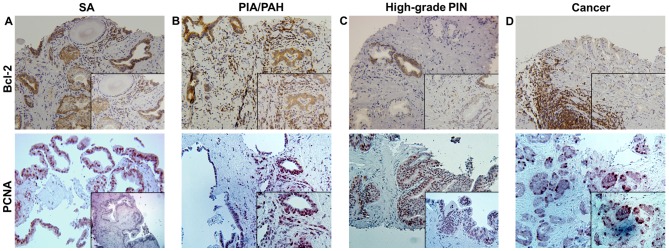
Figure 2.
Immunohistochemical staining for Bcl-2 and PCNA in needle-biopsy specimens within (A) SA, (B) PIA/PAH lesions, (C) high-grade PIN and (D) cancer. Bcl-2 is widely expressed in inflammatory and epithelial tissues of SA and PIA/PAH, with more intense staining in the basal epithelial cells near the areas of chronic inflammation, as well as predominantly in infiltrating immune cells. High expression of PCNA is observed in the proliferating epithelium in high-grade PIN and cancer. Magnification, ×20 (inset, ×40). PCNA, proliferating cell nuclear antigen; SA, simple atrophy; PIA, proliferative inflammatory atrophy; PAH, post-atrophic hyperplasia; PIN, prostatic intraepithelial neoplasia.

