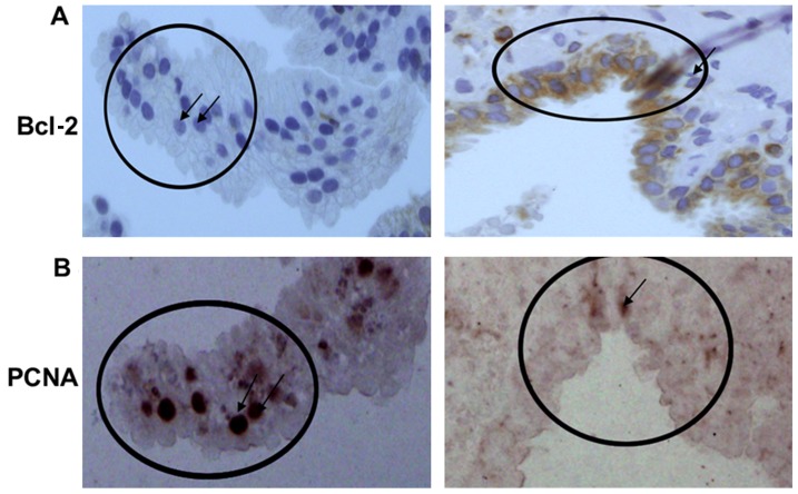
Figure 4.
Needle-biopsy specimens immunostained for Bcl-2 and PCNA in serial sections. Left and right panels demonstrate serial section from 2 different prostate needle biopsy specimens. (A) Epithelial cells show weak Bcl-2 staining (left panel) and heavy cytoplasmic staining for Bcl-2 (right panel). (B) Needle-biopsy specimens immunostained for PCNA within the serial sections of the same specimen. Intense PCNA in cells which do not express Bcl-2 (left panel) whereas little-to-no expression of PCNA is observed in cells expressing Bcl-2 (right panel). Arrows showing cells expressing high PCNA but not Bcl-2, and vice versa. PCNA, proliferating cell nuclear antigen.

