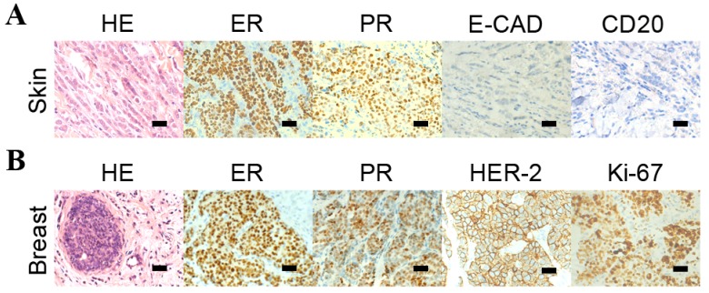
Figure 1.
HE staining and immunohistochemistry analysis of the breast tumors, the skin nodules. (A) HE staining revealed the malignant cells in the skin nodules and the immunohistochemistry analysis indicated that the cells from the skin nodules were positive for ER and PR and negative for E-CAD and CD20. (B) HE staining identified the malignant cells of the primary breast tumor and the immunohistochemistry analysis detected that the cells from the breast tumor were positive for ER, PR, HER-2 and Ki-67.

