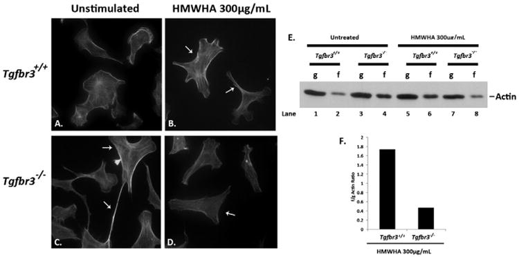
Fig. 2.

Defective actin dynamics in Tgfbr3−/− epicardial cells in the presence of HMWHA. (A–D) Detection of filamentous actin in Tgfbr3+/+ epicardial cells without (A) and with HMWHA 300 μg/mL stimulation for 60 min (B). Detection of filamentous actin in Tgfbr3−/− epicardial cells without (C) and with HMWHA 300 μg/mL stimulation (D) for 60 min. (E) Globular (g) or filamentous (f) actin detection by Western blot following ultracentrifugation of lysates from vehicle incubated and HMWHA-stimulated epicardial cells. (F) Representative graph of f/g actin ratios for HMWHA 300 μg/mL stimulated Tgfbr3+/+ and Tgfbr3−/− epicardial cells.
