Fig. 4.

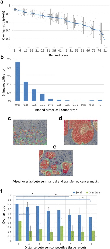
Overlap between manual and transferred cancer masks. (a) Error in alignment of transferred and ground truth masks. The overlap ratio was calculated as described in Materials and Method and plotted on the y-axis. The mean represents the average overlap in 3–5 images from each case and the standard deviation is indicated by the error bars. Cases are ranked in descending order of overlap ratio along the x-axis. (b) Distribution of cell count error. The difference of tumor cell counts underneath the transferred versus ground truth tumor masks was determined. 358 images were assigned to bins based on the error of tumor cell counts (see Materials and Methods) underneath the transferred tumor mask. (c-e) Visual demonstration of the error inflicted by the mask transfer. The transferred mask is outlined in green and the ground truth mask in red. Notice the association between the magnitude of error and the growth pattern by comparing the glandular growth pattern in (c) and (e) and solid growth pattern in (d). (e) shows a close-up of an area in (c). (f) Comparison of the error of mask transfer in solid (blue) versus glandular (green) regions of invasive breast cancer. Serial sections from 3 tumor blocks of invasive breast cancer were stained with Pan-CK. Solid and glandular areas were separately annotated and the masks transferred between slides. The x-axis indicates the distance between serial sections (unit length = 4 μm) from the same block and the y-axis demonstrates the overlap ratio of the transferred versus hand-annotated masks within solid versus glandular regions of invasive breast cancer. The difference in the overlap between solid and glandular regions was significant (* = p < 0.05)
