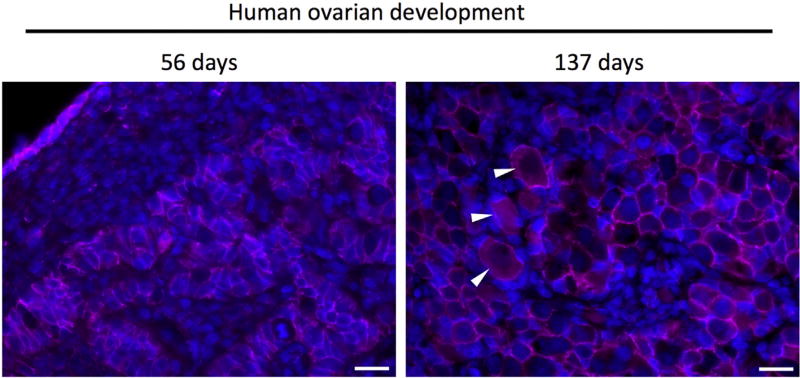
Fig. 1.
Immunofluorescent micrographs of human ovarian tissue during development (56 days, 137 days) and from reproductive-age ovarian tissue reveals break down of the germ cell nests and formation of primordial follicles. At 56 days of development, PGCs/oogonia cluster in cords, segregated from somatic cells. Subsequently, germ cell nests begin to breakdown (shown here at 137 days of development) to create primordial follicles (white arrows in center image) consisting of an oocyte surrounded by several squamous pregranulosa cells. In the adult human ovary, primordial follicles persist as an oocyte surrounded by a single layer of pre-granulosa cells. Scale bar = 20 microns. Immunofluorescence staining for beta-catenin (pink) and counterstained with Hoechst to stain nuclei (blue). (For interpretation of the references to colour in this figure legend, the reader is referred to the web version of this article.)

