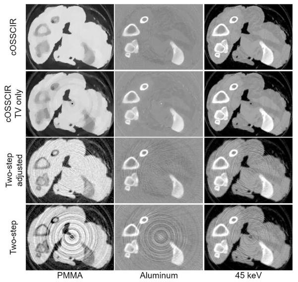
Fig. 11.
Experimental results. PMMA and aluminum basis images of the tissue specimen reconstructed by the proposed cOSSCIR method, cOSSCIR with only a TV constraint (no spectral-response scaling) and the two-step approach that assumed empirically estimated spectra with and without scaling correction. Images representing the 45 keV image are also displayed for each reconstruction approach. The display windows are [−0.1, 0.1] for the PMMA images, [−0.3, 0.3] for the aluminum images, and [0, 0.4] for the 45-keV image. The basis map values are unitless while the 45 keV images are in units of cm−1.

