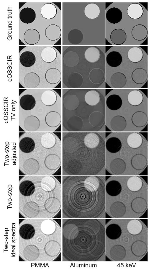
Fig. 6.
Experimental results. PMMA and aluminum basis images reconstructed by the proposed method (labeled as “cOSSCIR”), cOSSCIR algorithm with only a TV constraint (i.e., no spectral-response scaling, labeled as “cOSSCIR TV only”) and the two-step approach that assumed empirically estimated spectra (labeled as “Two-step”), estimated spectra with scaling correction (labeled as “Two-step adjusted”), and ideal spectra (labeled as “Two-step ideal spectra”). Images representing the 45 keV image are also displayed for each reconstruction approach. A ground-truth phantom image is also displayed. The display windows are [−0.1, 1.5] for the PMMA images, [−0.1, 0.2] for the aluminum images, and [0, 0.6] for the 45-keV image. The basis map values are unitless while the 45 keV images are in units of cm−1.

