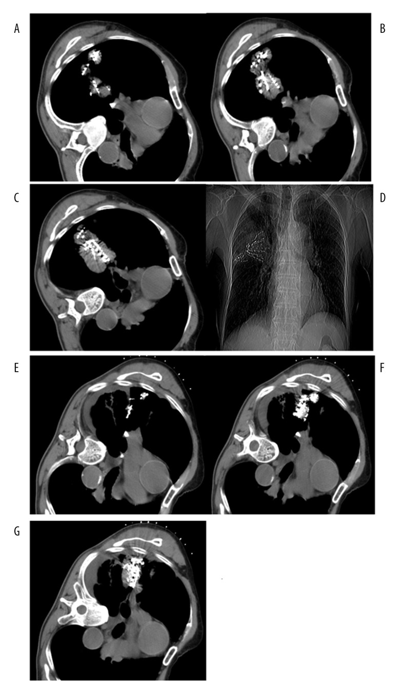
Figure 3.
(A–G) A 51-year-old male with poorly differentiated squamous carcinoma of the right lung. (A–C) Two work casings were used to implant seed from different directions in a large lesion; seed distribution was satisfactory on multiple planes. (D) X-ray radiography showed that there were 2 fan-shaped seed images in the lesion. (E, F) CT images at 6 months after seed implantation showed obviously reduced lesions and seed aggregation was found at the corresponding level.

