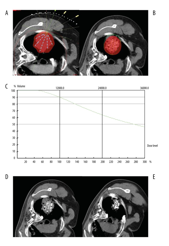
Figure 5.
(A–E) A 77-year-old male patient with poorly differentiated adenocarcinoma of the right lung. (A) According to TPS preoperative planning, seeds showed a fan-shaped distribution in single plane, and red color presented the coverage area of prescription dose. (B) TPS postoperative verification showed that seed distribution was different from that of preoperative plan; the prescription dose did not cover the whole lesion, and there was a dose cold-area near the pleura. (C) Dose curve of TPS postoperative verification showed 90% of lesion volume was covered by 120GY seed dosage. (D) CT image immediately after seed implantation displayed a fan-shaped distribution of seeds. (E) CT image at 2 months after seed implantation showed obvious reduction of the lesion and seed aggregation. Seeds were supplemented through implantation needle in the dose cold-area near the pleura.

