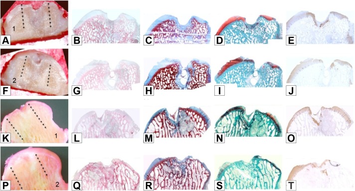
Figure 5.
Untreated group: macro and histological observations of 2 condyles, sectioned in half (number 1 in images A and K: right hemicondyle; number 2 in images F and P: left hemicondyle).
Notes: Macroscopic appearance: A, F, K, P. Histological appearance: B, G, L, Q haematoxylin and eosin staining; C, H, M, R azan-mallory staining; D, I, N, S safranine-O staining; E, J, O, T type II collagen immunostaining. Magnification ×20.

