Fig. 1.
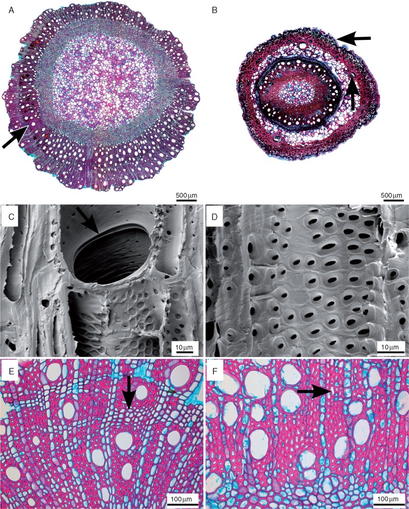
Wood anatomical sections of Nepenthaceae. Transverse light microscope sections (A, B, E, F) along with radial (C) and tangential (D) scanning electron microscopy surfaces of Nepenthes wood. (A) Nepenthes khasiana, mature stem (bark detached) showing wood with an indistinct growth ring (arrow). (B) Nepenthes muluensis, entire juvenile stem with pronounced cuticle (horizontal arrow) and lignified areas in both the outer stem area (cortex) and the inner stem part (wood and outer pith region); the vertical arrow points to the vascular bundle in the cortex. (C) Nepenthes tobaica, bordered, simple perforation plate with rim (arrow). (D) Nepenthes smilessi, alternate intervessel pits. (E) Nepenthes smilessi, tendency to form banded axial parenchyma (arrow). (F) Nepenthes edwardsiana, diffuse-in-aggregates axial parenchyma (arrow).

