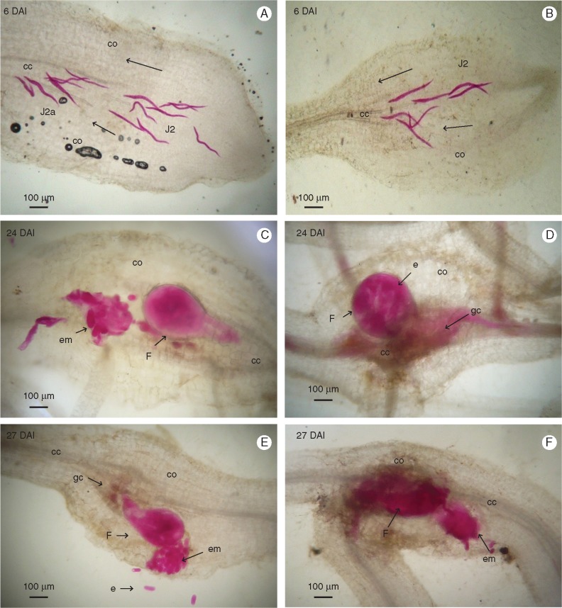
Fig. 1.
Histopathological examination of root sections of Musa acuminata genotypes CAV (left) and 4279-06 (right) infected with Meloidogyne incognita. Root sections (10 μm) were observed under brightfield microscopy following staining with acid fuchsin. (A, B) Second-stage juveniles (J2) migrating inside the root tip. (C) Adult females with egg masses released in the cortical zone. (D) Adult female attached to the central cylinder. (E, F) Adult females with egg masses released in the cortical zone. DAI, days after inoculation; cc, central cylinder; co, cortex; em, egg mass; e, egg; F, female; gc, giant cell.

