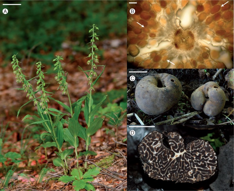
Fig. 1.
(A) Epipactis neglecta at forest site 9 in the Nördliche Frankenalb in July 2009. Scale bar = 5 cm. Image courtesy of Florian Fraaß. (B) Light micrograph showing a transverse section of a root of Epipactis neglecta. Fungal colonization is visible as exodermal, outer and inner cortex cells filled with fungal hyphae, indicated by white arrows. Scale bar = 100 µm. (C) Ascocarps of Tuber excavatum. Scale bar = 1 cm. (D) Cross-section of an ascocarp of Tuber brumale. Scale bar = 1 cm.

