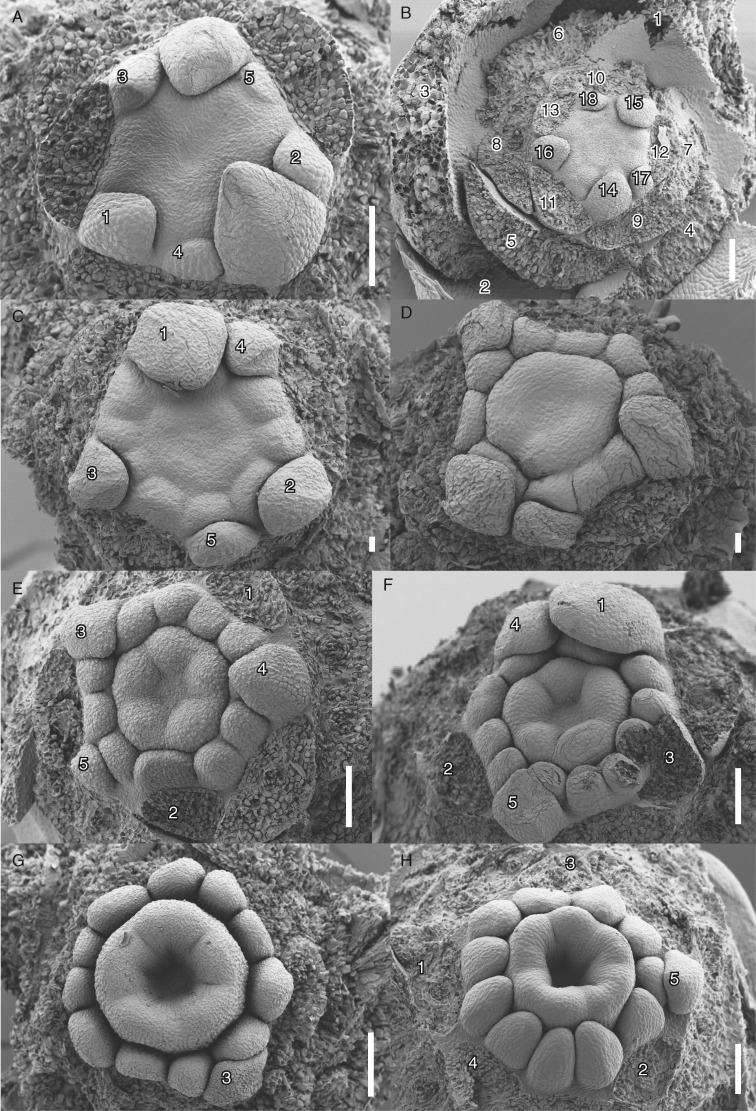
Fig. 3.
Floral development of Berberidopsis beckleri after stamen and carpel initiation. (A) Appearance of peripheral stamen protuberances and development of a central depression. (B) Similar stage with 18 organs, showing all bracts and tepals. (C) Differentiation of stamens at the floral periphery and appearance of carpels. (D) Upward growth of gynoecium and development of stamens. (E, F) Differentiation of five hemispherical carpels. (G) Apical view showing the development of a central gynoecial cavity. (H) Similar lateral view with progressive differentiation of the anthers. Numbers indicate order of initiation of tepals and bracts; the upper five petaloid tepals are numbered separately, except in (B), where they are shown as a full sequence of all tepals. Scale bars = 100 µm, except (C, D) = 20 µm.

