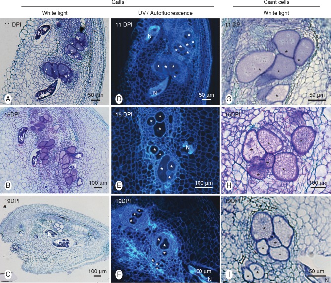
Fig. 2.
Histological analysis of galls induced by Meloidogyne graminicola in resistant rice (Oryza glaberrima TOG5681) roots. Gall cross-sections (10 µm) obtained at 11, 15 and 19 dpi were observed under UV light (middle panel) or were stained with toluidine blue and observed with white light (left and right panels). (A, B, C) Multiple nematode feeding on giant cells within the same gall. Note degraded nematode bodies and empty giant cells at 19 dpi (C). (D, E, F) Autofluorescence of sectioned galls. Note the absence of fluorescence accumulation around giant cells and nematodes. (G, H, I) Time course of giant cell degradation. (G) 11 dpi: dense cytoplasm and a large number of nuclei encircled by numerous neighbouring parenchymatic cells. (H) 15 dpi: vacuolated cytoplasm. (I) 19 dpi: degraded, devoid of cytoplasm and nuclei. Asterisk, giant cell; N, nematode.

