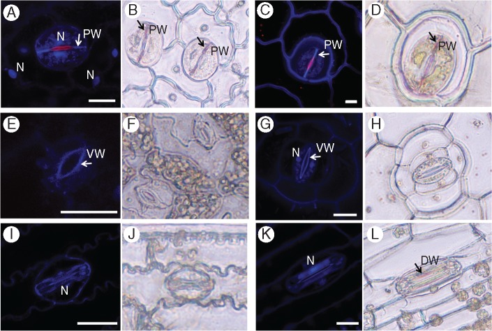
Fig. 7.
Lignins and phenolic compounds in stomatal guard cells. Lignin (blue) and phenolic compounds (red) autofluorescence was observed in leaf fragments using confocal microscopy (A, C, E, G, I, K) and by phloroglucinol stain in epidermal peels (B, D, F, H, J, L). (A, B) Asplenium – note the phenolic compound autofluorescence in the nuclei and red autofluorescence of the ventral wall; (C, D) Platycerium – note the red autofluorescence of the ventral wall (white arrow); (E, F) Arabidopsis; (G, H) Commelina; (I, J) Sorghum; (K, L) Triticum. N, nucleus; PW, polar end-wall; VW, ventral wall; DW, dorsal wall. Scale bars = 20 µm.

