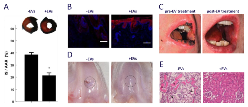
Figure 1. Clinical and pre-clinical studies using EVs derived from mesenchymal stem cells.
(a) Exosomes are well known to be effective in myocardial tissue repair after ischemia-reperfusion injury; in this mouse model, infarct size (IS, stained white) as a proportion of area-at-risk (AAR, stained red) was reduced from 39 ± 2% to 21 ± 2% (t = 1 day). Images were adapted from Arslan et al.68 and reproduced with permission from Elsevier. (b) Exosomes were used to promote wound healing in a rat model of skin deep second degree burn injury. Immunostaining of CK19 expression (red) along with Hoechst stain (blue) showed re-epithelization at the wound area for rats treated with exosomes (t = 2 weeks, scale bars = 200 μm). Images were reproduced under creative commons licence from Zhang B et al. (2015) Stem Cells doi:10.1002/stem.1771.86 (c) Exosomes were used in a clinical study to reduce pro-inflammatory cytokine response and alleviate the symptoms of therapy-refractive graft-versus-host disease. Images were adapted from Kordelas et al.109 and reproduced with permission from Nature Publishing Group. (d) Exosomes have also been used to enhance in vivo cartilage repair in 1 mm deep osteochondral defects created on the trochlear grooves of distal femurs of adult rats (t = 6 weeks). Images reproduced under creative commons licence from Zhang S et al. (2016) Osteoarthritis and Cartilage doi:10.1016/j.joca.2016.06.022.82 (e) Microvesicles have been shown to provide protection against tubular injury in an acute kidney injury mouse model. Here, cisplatin was used to induce intra-tubular casts (asterisks) and tubular necrosis (arrows), which was alleviated with multiple injections of microvesicles (t = 4 days, magnification = 200X). Images were reproduced under open access from Bruno S et al. (2012) PLoS ONE 7(3) doi:10.1371/journal.pone.0033115.77

