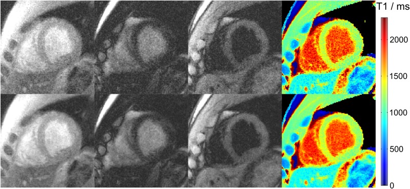
Figure 4.
Cardiac images and T1 maps (top) without and (bottom) with spatial filtering. Single-shot inversion recovery FLASH was performed at 43 ms temporal resolution (19 spokes) for a duration of 3 s. The images (magnified views) refer to an early time point after inversion, nulling of myocardial tissue and nulling of blood signal, respectively.

