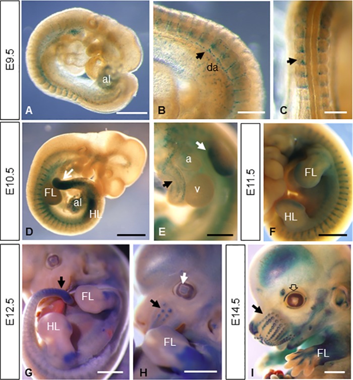
Fig 2. Whole-mount β-gal stained embryos from E9.5 to E14.5.

(A-C) E9.5 embryos (n = 7) show reporter expression in the allantois (A), the intersomitic arteries sprouting from the dorsal aorta (arrow in B) and in the posterior portion of the somites (arrow in C). (D-F) This vascular pattern of expression persists during somite development as shown in D and F. Other embryonic tissues like the limb buds, the mesenchymal tail tip (arrow in D and G), the allantois, the atrium and aorta (E) show β-gal staining at E10.5 (n = 5) and E11.5 (n = 3). At later stages of development, expression persists in the limbs (G-I) and appears in the primordium of follicle of vibrissa (arrows in H and I), lens (white arrow in H), eyelid (empty arrow in I), nose, pinna of the ear and brain (n = 6 for E12.5 and E14.5 embryos). Abbreviations: a, atrium; al, allantois; da, dorsal aorta; FL, forelimb; HL hindlimb; v, ventricle. Scale bars: 1 mm (D, F-I), 500 μm (A, E) and 250 μm (B, C).
