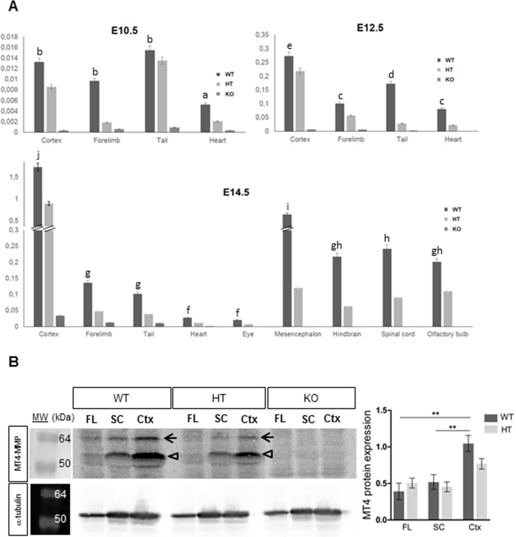
Fig 4. Real-time PCR and western blot analysis of Mt4-mmp expression in mouse embryonic tissues.
(A) Real-time PCR analysis was performed using cDNA prepared from E10.5, E12.5 and E14.5 WT, HT and KO mouse embryonic tissues. Mt4-mmp expression increases as development proceeds and particularly, high RNA levels were detected at E14.5 in distinct brain regions as the cerebral cortex, olfactory bulb, mesencephalon, hindbrain and spinal cord. Mt4-mmp RNA levels of were normalized to the housekeeping GAPDH levels and relative to expression levels in the adult cerebral cortex. Data shown are representative of three independent experiments and expressed as the mean ± SEM. In all cases, mRNA levels were significantly reduced in the HT compared to the WT tissues at the distinct embryonic stages analysed. Different letters indicate significant differences (p< 0.05) among distinct tissues of the same embryonic stage by ANOVA and post-hoc analysis (SNK). (B) Total cell lysates from forelimbs (FL), spinal cord (SC) and cerebral cortex (Ctx) of E14.5 WT, HT and KO embryos were analysed by western blotting with the anti-MT4-MMP antibody and anti-α-tubulin antibody for loading control. Specific bands correspond to the precursor of the GPI-anchored form (63 kDa, closed arrow) and the GPI-anchored latent and active forms (55 and 50 kDa, opened arrow) of MT4-MMP. Quantification of Western blot shows significant higher levels of protein expression in the cerebral cortex compared to the spinal cord and the forelimb of WT embryos. MT4-MMP protein levels were significantly decreased in the HT cerebral cortex compared to the WT embryos and were totally absent in all KO embryonic tissues analysed (**, indicates significant differences at p< 0.01).

