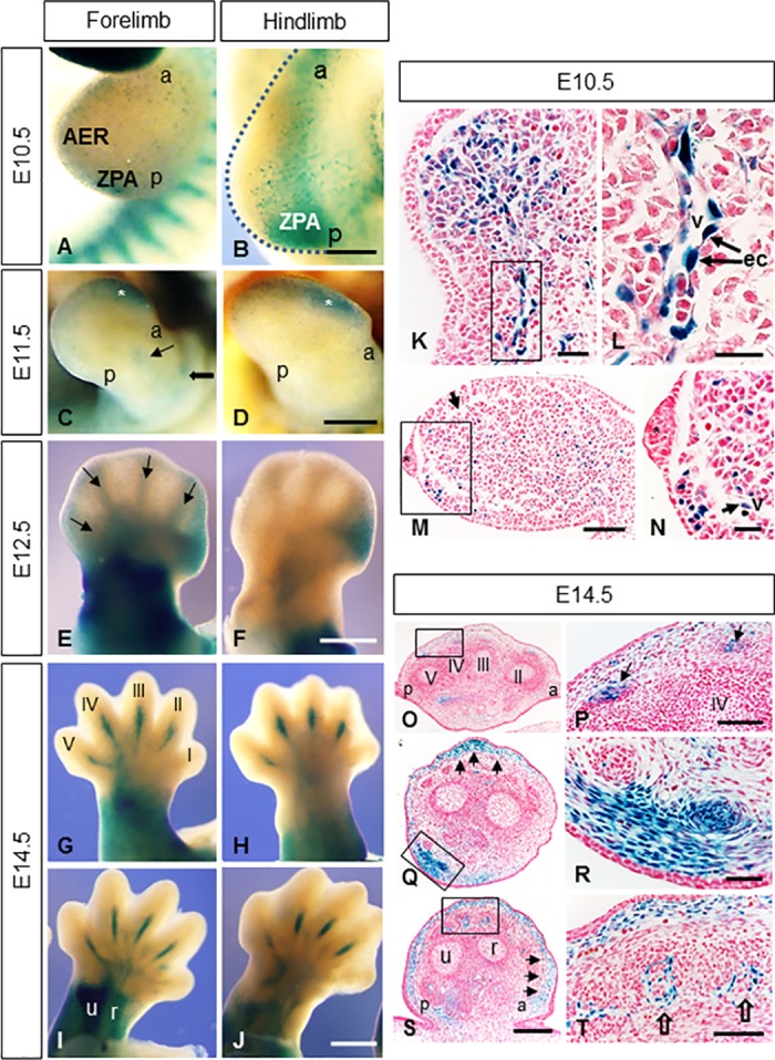
Fig 5. Mt4-mmpLacZ/+ distribution during mouse limb development.

Mt4-mmpLacZ+/- expression pattern was studied in whole mount limbs at distinct embryonic stages. (A, B) Distal (A) and proximal regions (B) of the limb buds as well the ZPA region show β-gal staining at E10.5. (C, D) Mt4-mmp expression was detected in distal regions of the hand and footplate (asterisks in C,D) as well as in the developing stylopodial and zeugopodial segments (thick and thin arrows in C). (E-J) β-gal staining from E12.5 to E14.5 appears in the stylopodium, zeugopodium and proximal regions of the handplate (E, G-J). In addition, anterior mesenchymal expression is observable in the E12.5 footplate (F). Mt4-mmp expression was first observed in digits at E12.5 (arrows in E) evolving to a pattern restricted to metacarpals and metatarsals of digits II, III, IV and V, either in dorsal (G, H) and ventral side (I, J) at E14.5. (K-N) Paraffin cross sections though hindlimb (K, L) and forelimb (M, N) showed β-gal positive cells in the mesenchyme (arrow in N) and in endothelial cells (ec) of blood vessels entering the limbs (L). The asterisk corresponds to the AER. (O-T) E14.5 limb crossed-sections from distal (O, P) to proximal (Q-T) levels evidence β-gal staining in the tendon blastemas (arrows in P), in subepidermal mesenchymal cells (arrows in Q, S) and in ventral muscle blocks (R) as well as in blood vessels entering the muscles (empty arrows in T). In all sections dorsal is up; ventral is down. Abbreviations: a, anterior; AER, apical ectodermal ridge; ec, endothelial cells; p, posterior; r, radius; u, ulna; v, blood vessel; ZPA, zone of polarizing activity. Scale bars: 500 μm (C-J), 250 μm (A, B, O, Q, S), 100 μm (M, P, T), 50 μm (K, R) and 25 μm (L, N).
