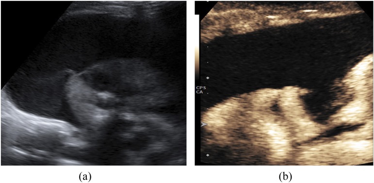
Figure 4.
A 4-year-old male with pneumonia complicated with empyema: B-mode ultrasound (a) is showing lung consolidation and the presence of an anechoic parapneumonic effusion. Contrast-enhanced ultrasound (b) is demonstrating the enhancement of the consolidated lung parenchyma and has accurately delineated the empyema, which showed no enhancement.

