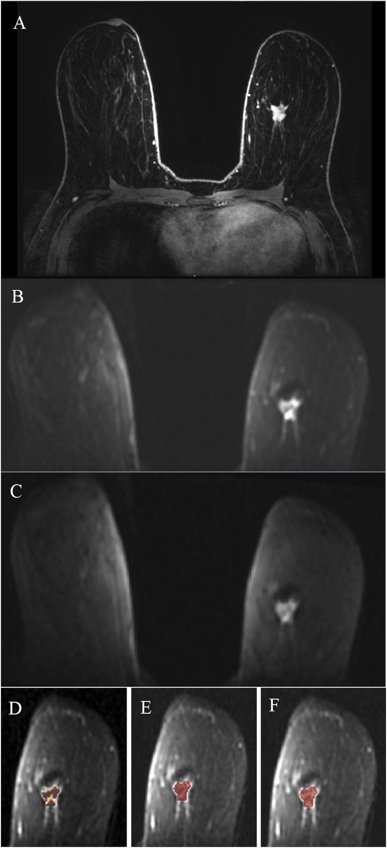
Figure 5.
Invasive ductal carcinoma in the left breast in a 42-year-old female: the mass is visible on dynamic contrast-enhanced MRI (a) and intravoxel incoherent motion (IVIM) images (b, c) at b = 0 s mm−2 and b = 500 s mm−2. IVIM protocol b-values = 0, 10, 192 and 500 s mm−2; d, e and f are showing the IVIM parameter maps for f, D and D*, respectively. Histogram analysis has gave median values of f = 22%, D = 147 × 10−6 mm2 s and D* = 7773 × 10−6 mm2 s.

