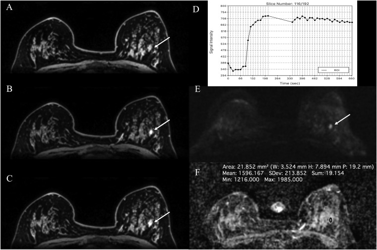
Figure 7.
MRI of the breast at 3.0 T: fibroadenoma in a 42-year-old female at 2 o'clock in the right breast—the round and partly irregularly marginated mass [(a) pre-contrast media, (b) initial and (c) delayed enhancement] is demonstrating a homogeneous fast/washout contrast enhancement (d) and has been classified as breast imaging-reporting and data system (BI-RADS) 4 on dynamic contrast-enhanced MRI. On diffusion-weighted imaging (e), the apparent diffusion coefficient values (1.596 × 10⁻³ mm2/s) (f) were well above the threshold for malignancy, thus allowing an accurate classification as a benign finding (BI-RADS 3—probably benign) with the BI-RADS-adapted reading. ROI, region of interest.

