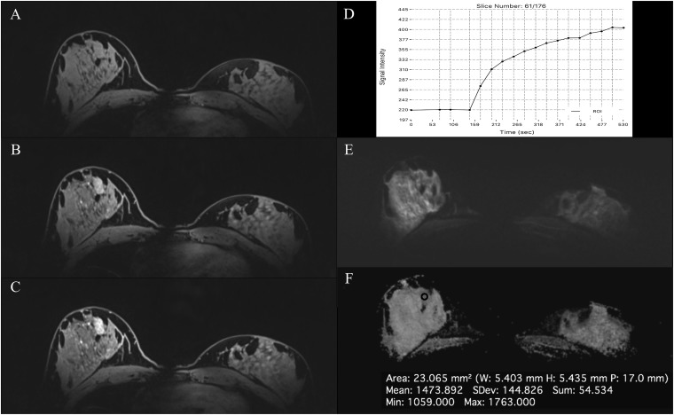
Figure 8.
MRI of the breast at 7.0 T: invasive ductal carcinoma grade 3 and associated high-grade ductal carcinoma in situ in the right breast in a 36-year-old female—high-resolution dynamic contrast-enhanced MRI at 7.0 T is showing an irregular shaped, partly spiculated mass lesion retromammillary right (a) with fast/washout enhancement kinetics (b). Diffusion-weighted imaging is showing a restricted diffusivity (c) and low apparent diffusion coefficient values (0.68 × 10⁻³ mm2 s) (d), indicating high cellularity, which is indicative of tumour malignancy (breast imaging-reporting and data system 5—highly suggestive of malignancy). ROI, region of interest.

