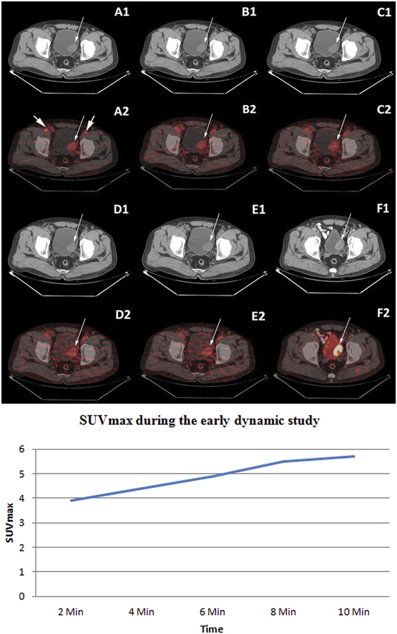
Figure 4.
Upper panel: staging fludeoxyglucose (FDG) positron emission tomography (PET)/CT displaying the non-enhanced transaxial CT (A1–E1) and corresponding early dynamic fused PET/CT images of the bladder (A2–E2). Increased flow of tracer in the bladder tumour [maximum standardized uptake value (SUVmax) 3.9 at 2 min] in the left posterolateral wall was noted in all the dynamic images (thin arrows) as well as in the bilateral external iliac vessels (thick arrows in A2). Intense FDG uptake (SUVmax 19.9) was noted in the bladder tumour on standard FDG PET/CT images after diuresis (F1: transaxial contrast-enhanced CT and F2: fused PET/CT image). Histopathology of bladder tumour following transurethral resection of the bladder tumour was of high-grade transitional cell carcinoma. Lower panel: time activity curve of lesion SUVmax in a high-grade bladder tumour during the early dynamic imaging of patient represented in the upper panel.

