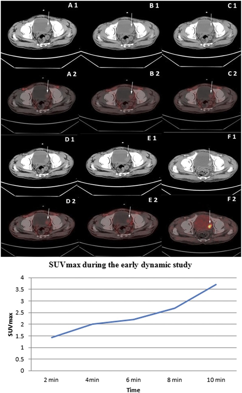
Figure 5.
Upper panel: staging fludeoxyglucose (FDG) positron emission tomography (PET)/CT displaying non-enhanced transaxial CT (A1–E1) and corresponding early dynamic fused PET/CT (A2–E2) images of the bladder. Mildly increased flow of tracer in the bladder tumour (SUVmax 1.44 at 2 min) in the left lateral wall was noted in all the dynamic images (thin arrow). Intense FDG uptake [maximum standardized uptake value (SUVmax) 9.8] was noted in the bladder tumour on standard FDG PET/CT images after diuresis (F1: transaxial CECT and F2: fused PET/CT image). Histopathology following transurethral resection of the bladder tumour revealed low-grade transitional cell carcinoma with no invasion of the detrusor muscle or the lamina propria. Lower panel: time activity curve of lesion SUVmax in a low-grade bladder tumour during the early dynamic imaging of patient represented in the upper panel.

