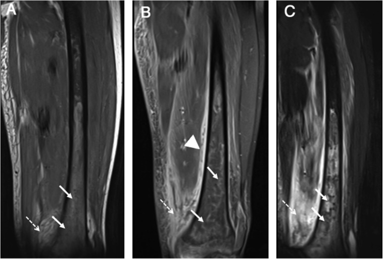
Figure 12.
Coronal T1 (a), coronal T1 post-contrast (b) and coronal short tau inversion-recovery (c) images of the femur demonstrating extensive marrow oedema with associated serpiginous and tubular marrow enhancement in the distal femoral metaphysis/diaphysis (solid arrows) with extensive associated periosteal reaction (arrowhead), adjacent soft tissue swelling and oedema (dashed arrows). These findings are characteristic of osteomyelitis.

