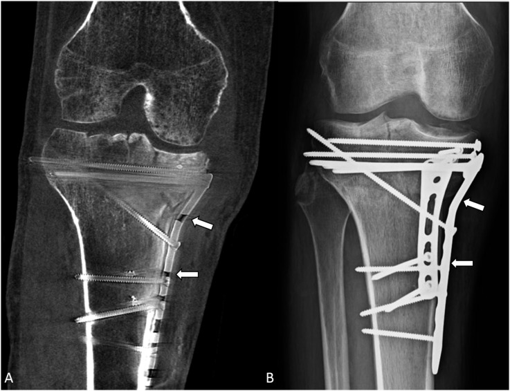
Figure 4.
A 48-year-old male with a history of a right unicondylar tibial plateau fracture followed by open reduction and internal fixation showing the comparison of metallic hardware contour between cone-beam CT (CBCT) (A) and radiograph (B). The metal plate (arrows) on the medial side of the tibia can be seen clearly in the radiograph; however, it is not as clearly seen on the CBCT image owing to artefacts.

