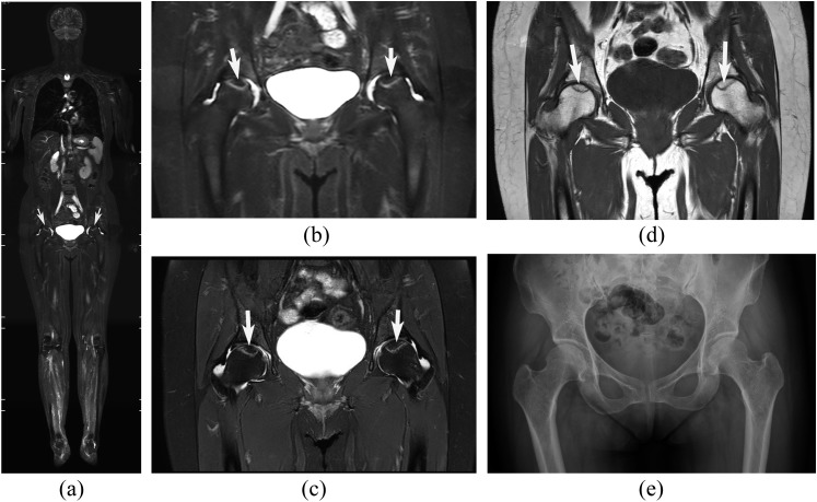
Figure 1.
Osteonecrosis of the bilateral femoral heads in a 50-year-old female with dermatomyositis. (a–e) Short-tau inversion recovery whole-body MRI (STIR-WBMRI) (a), the hip area of coronal STIR-WBMRI (b), corresponding regional hip MR T2 weighted spectral pre-saturation inversion recovery (T2W SPIR) (c), T1 weighted (T1W) images (d) and anteroposterior radiograph (e). The necrotic region (white arrows) is surrounded by a distinct high-intensity rim on STIR-WBMRI, T2W SPIR and is surrounded by a low-signal band on T1W. A high degree of consistency was demonstrated between STIR-WBMRI and regional hip MR on necrotic lesion size, shape and position. Radiography did not detect the osteonecrosis shown on STIR-WBMRI.

