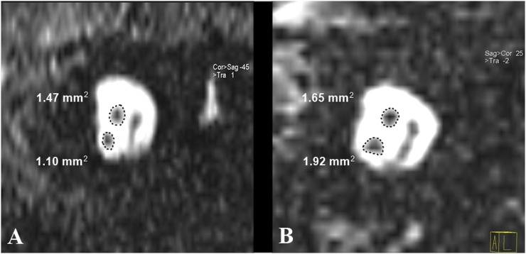
Figure 3.
Parasagittal reconstructed T2 weighted MRI from two patients (ROIs are schematically represented): one patient (a) from Group 1 with long-standing sensorineural hearing loss showing the small-sized cochlear nerve compared with the facial nerve and one patient (b) from Group 2 with cochlear nerve size larger than that of the facial nerve. cor, coronal; sag, sagital.

